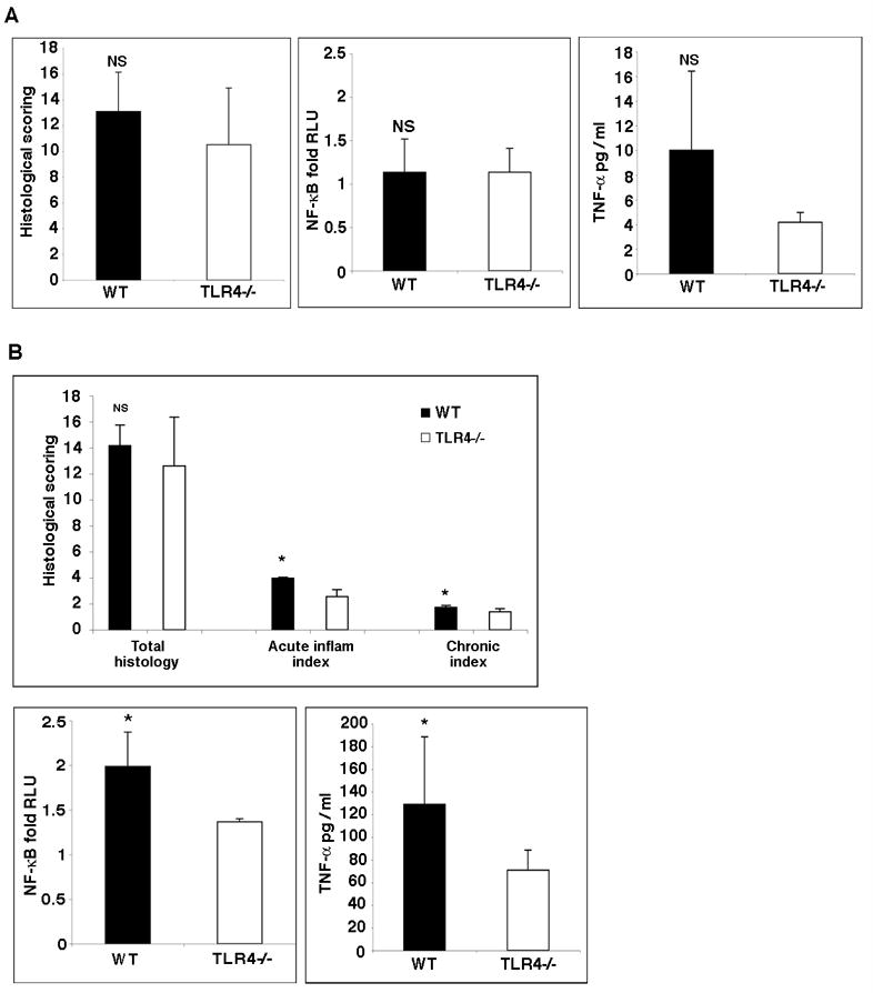
Figure 3. Comparison of inflammatory activity in TLR4−/− versus WT mice.

A. Histology scores in WT versus TLR4−/− mice at day 56 following AOM+DSS (left panel). Table 1 demonstrates the criteria used for scoring. The total histology score was similar in WT and TLR4−/− mice. NF-κB activation state (middle panel) and TNF-α secretion (right panel) in WT versus TLR4−/− mice at day 56 following AOM+DSS. There are no significant differences between WT and TLR4−/− mice in NF-κB activation or TNF-α production in the intestine.
B. Histology scores in WT versus TLR4−/− mice at day 7 following DSS. The acute and chronic sub-scores for the indices are shown. NF-κB activation state and TNF-α secretion in WT versus TLR4−/− mice after seven days of DSS treatment. TLR4−/− mice have significantly decreased NF-κB activation and TNF-α secretion.
