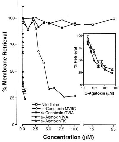
Figure 3.
The pharmacological characterization of the calcium influx required for membrane retrieval. Eggs were suspended in ASW and fertilized with sperm. After 3 minutes the eggs were incubated for 15 minutes in ASW containing the indicated concentration of ω-conotoxin MVIIC, an inhibitor of N-, P-, and Q-type voltage-activated calcium channels (○; all data points are mean ± SD, n = 3 except 0 μM was n = 6); nifedipine, an inhibitor of L-type voltage-activated calcium channels (□; all points are mean ± SD, n = 3); ω-conotoxin GVIA, a specific inhibitor of N-type voltage-activated calcium channels (■; all data points are mean ± SD, n = 6); ω-agatoxin IVA, a selective inhibitor of P-type voltage-activated calcium channels in this concentration range (●; all data points are mean ± SD, n = 6 except 2.5 and 5 nM which were n = 3);and ω-agatoxin TK, a selective inhibitor of P-type voltage-activated calcium channels in this concentration range (open triangles; all data points are mean ± SD, n = 3). The insert in B shows the ω-agatoxin IVA and ω-agatoxin TK dose–response curves on a logarithmic scale. All data points were normalized to controls lacking inhibitors.

