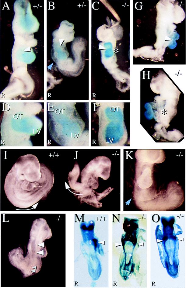
Figure 4.
KIF3A mutant embryos display morphological and asymmetrical defects. Blue staining in A–H is for the ventricle- specific MLC2v message (29). (A) A normal heterozygous 9.5 days p.c. embryo displaying proper cardiac looping. (B) A heterozygous mutant embryo exhibiting retarded cardiac looping. (C) A null mutant embryo displaying reversed cardiac looping. Arrowheads in A–C indicate direction of cardiac looping. (D–F) Closeup of hearts in A–C. (LV = Left Ventricle; OT = Outflow Tract). (G and H) Right and left side views of KIF3A null mutant embryo with reversed cardiac looping. (I and J) A wild-type embryo that underwent normal embryo turning and a KIF3A null embryo that failed to undergo embryonic turning. Arrow indicates the orientation of the embryo posterior. (K) KIF3A null mutant embryo exhibiting edema around the heart. Blue arrowheads in B and K indicate the membrane. (L) KIF3A null mutant embryo that has a neural tube closure defect. Large arrowheads point to open neural tube and small arrowheads point to closed neural tube. (M–O) In situ hybridization of 8.0-days p.c. embryos for Pitx2, ventral views. (M) Normal left side expression of Pitx2 in the lateral plate mesoderm in wild-type embryos. (N and O) Bilateral expression of Pitx2 in KIF3A null mutant embryos. Arrowheads point to strong lateral plate mesoderm staining.

