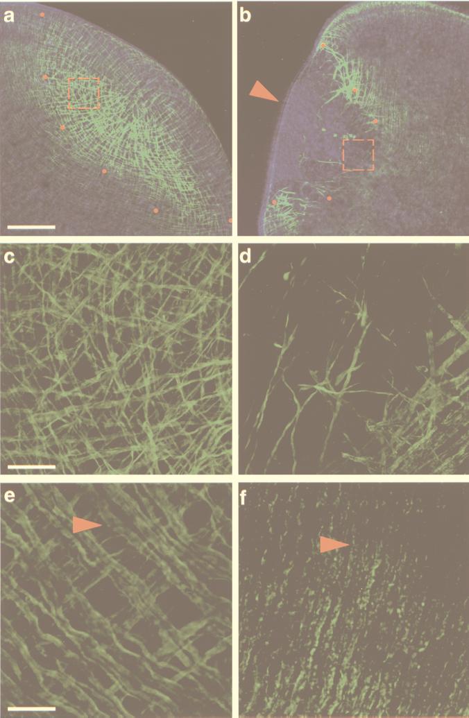
Figure 1.
Effects of myosin dsRNA injection in the planarian S. mediterranea. Confocal projections of body-wall musculature visualized with an anti-myosin heavy chain mAb (21). (a, c, and e) Control animals injected with water. (a and c) The normal dynamics of body-wall muscle regeneration. (e) The intact, terminally differentiated body-wall musculature. (b, d, and f) Myosin dsRNA-injected animals. Note the lack of appropriately regenerated muscle in b. (a and b) Confocal projections of 16 optical sections (1.86-μm intervals). Red dots demarcate the proximal margin of the blastema, and the red, dashed squares denote the areas magnified in c and d. The red arrowhead in b points to the blastemal epithelium imaged by superposing the phase-contrast image on the confocal projection. (c) The regenerating musculature with longitudinal (from left to right), circular (from top to bottom), and diagonal fibers within the blastema. (d) The few disorganized muscle fibers present in the proximal boundary of the blastema are shown with the preexisting body-wall musculature (lower right-hand corner). Note the punctate appearance of these fibers as compared with those shown in c. (c and d) Confocal projections of eight optical sections (0.65-μm intervals). (e and f) Confocal projections of eight optical sections (0.45-μm intervals) of the preexisting body-wall musculature in control and dsRNA-injected animals, respectively. Red arrowheads point to circular fibers. [Bars = 100 μm (a and b), 20 μm (c and d), and 10 μm (e and f).]

