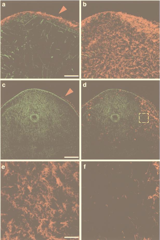
Figure 2.
Myosin and α-tubulin dsRNA injections in S. mediterranea. Confocal projections of body-wall musculature and ventral ciliated epithelium visualized with anti-myosin heavy chain and anti-acetylated tubulin (α-AcTub) mouse mAbs, respectively. The tubulin signal was pseudo-colored in red and was separated from the musculature signal by projecting only optical sections corresponding to the blastema’s ventral surface. (a) Projection of the complete subepithelial musculature (in green) in a 7.5-day cephalic blastema of an animal injected with myosin dsRNA (16 optical sections, 1.2-μm intervals). Note the disorganization of the musculature. The epithelium surrounding each of the optical sections and recognized by the α-AcTub antibody was pseudo-colored in red (arrowhead) and appears normal. (b) Superposition of the projected ventral epithelium (eight optical sections, 0.48-μm intervals) on the confocal projection shown in a. (c) Projection of the complete subepithelial musculature (in green) in a caudal blastema of an animal injected with α-tubulin dsRNA (16 optical sections, 1.41-μm intervals). No muscle defects are observed. The circular aperture is the normally regenerated pharyngeal opening. As in a, the epithelium recognized by the α-AcTub antibody was pseudo-colored in red (arrowhead). (d) Superposition of the projected ventral epithelium (eight optical sections, 0.35-μm intervals) on the confocal projection shown in c showing the disruption of the ciliated epithelium. (e) Confocal projection of 10 optical sections (0.21-μm intervals) of a normal (water-injected) caudal blastema. (f) The area demarcated by the yellow, dashed square in d shown at higher magnification. [Bars = 50 μm (a and b), 100 μm (c and d), and 20 μm (e and f).]

