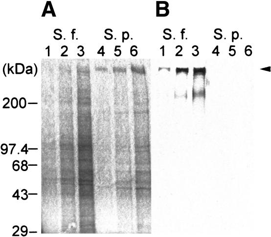
Figure 1.
Species specificity of Sf-H2. (A) Coomassie blue staining of purified VEs from S. franciscanus and S. purpuratus separated on a 4%–20% SDS-PAGE gel. (Lanes 1–3) S. franciscanus VE. (Lanes 4–6) S. purpuratus VE. (B) Western blot analysis of VE from S. franciscanus and S. purpuratus separated on a 4%–20% SDS-PAGE gel (shown in A) using anti-Sf-H2 antibody. The arrow highlights the ∼350-kD protein detected by the anti-Sf-H2 antibody. (Lanes 1–3) S. franciscanus VE. (Lanes 4–6) S. purpuratus VE. Lanes 1 and 4 are 2 μg of VE protein, lanes 2 and 5 are 5 μg of VE protein, and lanes 3 and 6 are 10 μg of VE protein.

