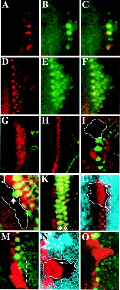
Figure 2.
The requirement of ato and da in MAPK activation. Early third-instar larval eye discs of +/+ (A–F), and ato1/ato1 (G and H) showing Ato (red) and dp-ERK (green) expression. Mutant clone of ato1 (I) or dakx136 (J) across the MF lacking dp-ERK expression (green). The clone boundary (marked by white line) is revealed by the lack of β-galactosidase (I, red) or Da (J, red) expression. The thick arrow in J indicates a dp-ERK cluster that overlaps the clone boundary. (K) Nts eye disc incubated at restricted temperature showing two rows of Ato clusters (red and indicated by ∗) expressing dp-ERK (green). ato1 (L and M) or dakx136 (N and O) clones showing expansion of Ato expression (red in L–O) and lacking dp-ERK expression (green in M and O). The clone boundary (marked by white line) was revealed by lack of myc staining (blue in L and N). Within the clones, expansion of Ato-positive cells reaches the most posterior row of R8 precursors (marked by dotted white lines in L–O), except that near the margin Ato is still repressed in some cases (arrows in L and N).

