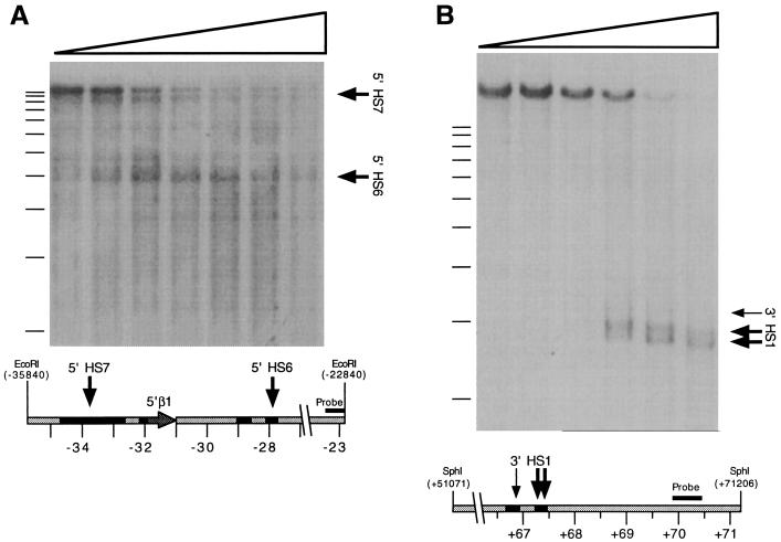
Figure 3.
DNaseI-HS mapping within the newly sequenced β-globin-flanking regions. (A) Mapping of human 5′ HS6 and 5′ HS7. A Southern blot of an EcoRI-digested DNase series prepared from human fetal liver was hybridized with a probe corresponding to an EcoRI/Eco0109I restriction fragment located at −23.4 to −22.8 kb. Arrows indicate DNaseI subbands evident on the autoradiogram and their corresponding positions on the human sequence represented below it. Filled regions on the diagram indicate significant (>50% identity) homology to mouse sequence, as shown in Fig. 2. (B) DNaseI-HSs located 3′ of mouse β-globin genes. A Southern blot of an SphI-digested DNase series prepared from phenylhydrazine-treated mouse spleen was hybridized with a probe corresponding to an AccI/NsiI restriction fragment located from +69.9 to +70.5 kb.

