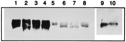
Figure 1.
Western blot analysis of hsp60 expression in mouse skin. Lanes 1–4 show hsp60 expression during rejection of BALB/c (H-2d) skin removed 10 days after transplantation to allogeneic NOD (H-2g7) mice. Lanes 5–8 show the expression of hsp60 in BALB/c skin transplanted 10 days earlier to syngeneic BALB/c mice. Lane 9 shows the spontaneous expression of hsp60 in untransplanted Eα.hsp60 transgenic NOD skin. Lane 10 shows the expression of hsp60 in untransplanted wild-type NOD skin.

