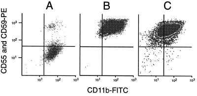
Figure 2.
(A) Granulocytes from PNH patient MSK11 stained with anti-CD55, anti-CD59, and CD11b-FITC, demonstrating a large PNH clone representing 80% of the cells. (B) Post-sort analysis of collected normal GPI(+) granulocytes from donor 8, demonstrating no residual PNH cells (compare with Fig. 1C). This confirms the ability of the cytometer to accurately sort a rare population. (C) A simulated experiment with PNH cells from patient MSK11 added to sorted GPI(+) cells from donor 1 at a frequency of 25 per million. The PNH population is clearly seen in the lower right quadrant, again confirming the ability of the instrument to identify this rare population. Events, most likely representing debris, also are seen in the lower left quadrant, demonstrating the usefulness of anti-CD11b for excluding these events.

