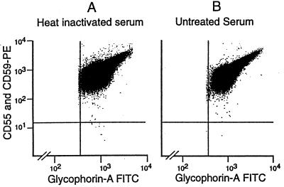
Figure 4.
PNH red blood cells in donor 8. Red cells are positively identified by light scatter characteristics and by gating to include glycophorin A(+) events. (A) Flow analysis of red blood cells incubated in a mock Ham test with heat-inactivated serum. PNH cells are clearly seen in the lower right hand quadrant at a frequency of eight per million. (B) A parallel analysis of red blood cells from donor 8 after incubation in the Ham test with untreated serum. The population in the lower right quadrant is reduced by a factor of 6, confirming that these PNH cells exhibit complement sensitivity.

