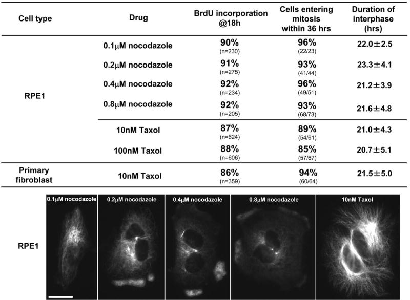
Figure 3.
Cell cycle progression of RPE1 and primary human fibroblasts in which the microtubule cytoskeleton has been diminished or augmented by 0.1-0.8μM nocodazole or 10-100 nM Taxol respectively. Upper portion shows the percent BrdU incorporation at 18 hours and the proportion of RPE1 cells or primary human fibroblasts entering a subsequent mitosis. The “duration of interphase” (mean, ± the standard deviation) is the time from shake off to the rounding up at the subsequent mitosis for cells continuously followed by time lapse video microscopy. The images show the extent of the diminishment/augmentation of the microtubule cytoskeleton in RPE1 cells continuously exposed to the indicated drugs and drug concentrations. The cells were fixed at 3 hours after shake off and immunostained for alpha tubulin. Fluorescence microscopy; Bar = 20μm.

