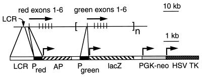
Figure 1.
Structure of a minimal human red/green visual pigment gene array in which the red pigment gene promotor controls transcription of an AP reporter and the green pigment gene promotor controls transcription of a β-gal reporter. The upper map shows the human X chromosome visual pigment gene array with the direction of transcription indicated by arrows and the six exons of each gene indicated by vertical lines (36–38). The LCR, defined by deletion mutations in blue cone monochromats (5) and by transgenic mouse experiments (6), is located between 3.1 and 3.7 kb 5′ of the start site of transcription of the red pigment gene. The lower map, at a 10-fold enlarged scale, shows the transgene construct which contains the following DNA segments, begining at the 5′ end: a BamHI–StuI fragment encompassing bases −4,564 to −3,009 5′ of the red pigment translation start site (LCR); a BamHI–NcoI fragment encompassing 496 bases 5′ of the red pigment gene initiator methionine codon (Pred, the red pigment gene promotor and 5′-untranslated region); a human placental AP-coding region (AP; ref. 8); the mouse protamine gene intron and 3′-untranslated region (open box; ref. 9); a PCR fragment (verified by sequencing) encompassing 496 bases 5′ of the green pigment gene initiator methionine codon (Pgreen, the green pigment gene promotor and 5′-untranslated region); an E. coli β-gal-coding region (lacZ; ref. 9); the mouse protamine gene intron and 3′-untranslated region (open box); a PGK-neomycin resistance cassette (PGK-neo; ref. 10); and a pMC1-HSV TK cassette (11, 12). The arrows indicate the start site and direction of transcription.

