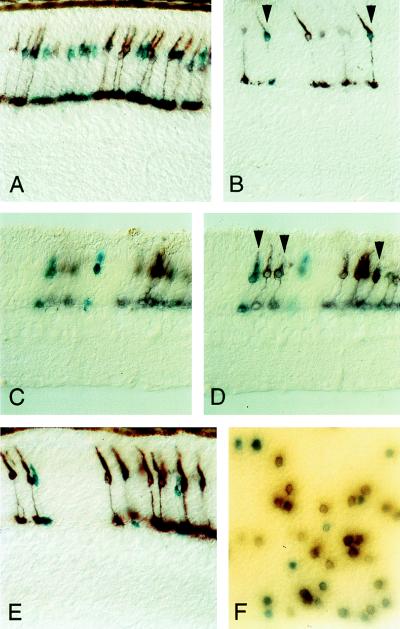
Figure 2.
Histochemical visualization of reporter enzyme activities in transgenic mouse retinae. Retinal sections 10–15 μm thick (A–E) and retinal flat mount (F) double-stained with X-Gal (blue reaction product) and X-phos/NBT (purple reaction product). In A–E, the full thickness of the retina is shown with the ganglion cell layer at the bottom of each panel. In A, B, and E, the tissue was processed with the retina and choroid attached; in C and D, the retina was dissected before histochemical processing. C and D show the same region of retina at different focal planes. Vertical arrows in B and D mark examples of double-stained cells: in D, the cell on the left has a greater level of X-Gal than X-phos/NBT staining, and the two other cells have greater levels of X-phos/NBT than X-Gal staining.

