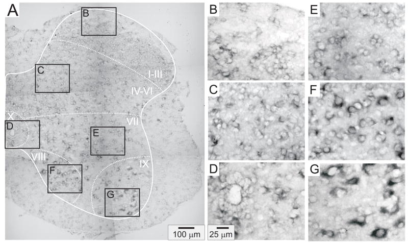
Figure 4.
D1 receptor distribution in the lumbar spinal cord. A: Photomontage of typical anti-sense D1 labeling. B–G: Enlargements of boxed regions in A for superficial dorsal horn (lamina I–III), deep dorsal horn (lamina IV-VI), central canal area (lamina X), intermediate gray (lamina VII), lamina VIII, and motor nuclei (lamina IX), respectively. In this and subsequent figures, the solid white line delineates spinal gray matter while dashed white lines approximate regions into laminae.

