
Figure 4.
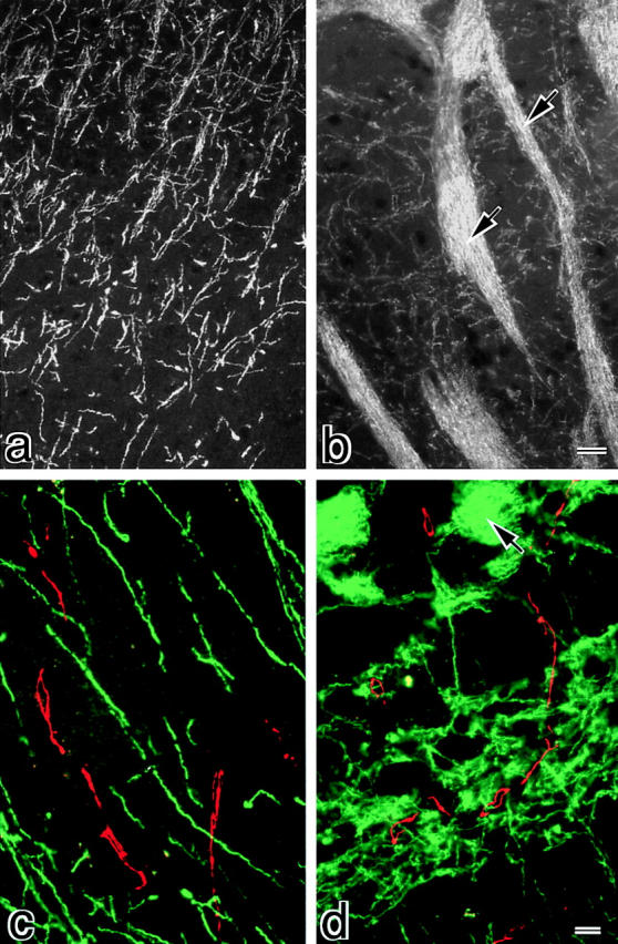
Distribution of claudin-11/OSP in mouse brain. Frozen sections of mouse brain, cortex region (a and c), and deeper region (b and d), were singly stained with anti–claudin-11/OSP pAb (a and b) or doubly stained with anti–claudin-11/OSP pAb (green in c and d) and anti-occludin mAb (red in c and d). In the cortex region (a and c), a large number of intensely stained linear structures was seen scattered in random directions, whereas in the deeper region (b and d) these claudin-11/OSP-positive structures were occasionally arranged in a parallel manner to form thick bundles (arrows). These claudin-11/OSP-positive linear structures did not overlap with occludin-positive endothelial TJs (c and d). Bars: (a and b) 10 μm; (c and d) 5 μm.