
Figure 8.
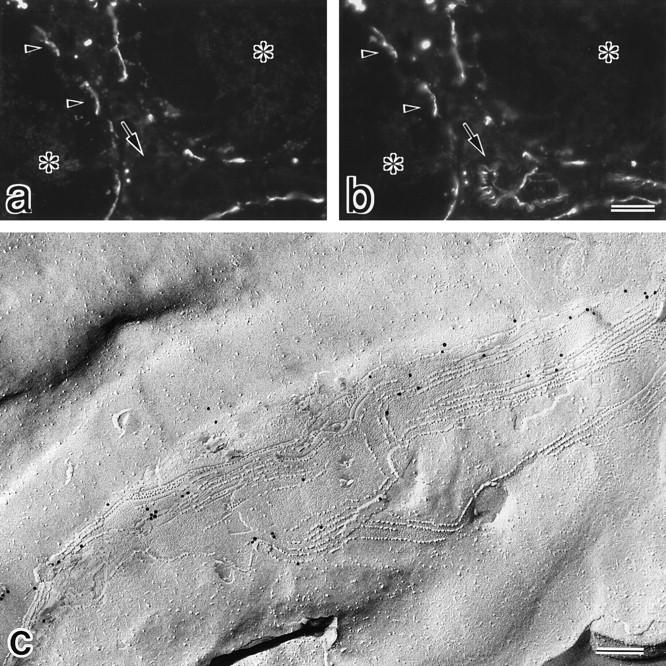
Subcellular localization of claudin-11/OSP at TJ strands in Sertoli cells. (a and b) Frozen sections of mouse testis were doubly stained with anti–claudin-11/ OSP pAb (a) and anti-occludin mAb (b). Both claudin-11/OSP and occludin were concentrated and precisely colocalized in a linear fashion at the most basal region of lateral membranes of adjacent Sertoli cells (arrowheads). Note that vascular endothelial cells were stained positively for occludin but were negative for claudin-11/ OSP (arrow). Asterisks, centers of seminiferous tubules. (c) Mouse testes were quickly frozen without chemical fixation, and then processed for freeze-fracture. Freeze-fracture replicas were labeled with anti–claudin-11/OSP pAb. Characteristic Sertoli TJs were exclusively labeled with the pAb (10-nm gold particles). Bars: (a and b) 25 μm; (c) 200 nm.