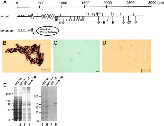
Figure 2.
Expression of the NH2-terminal sequence of Ten-m1 linked to an AP module in HEK 293 cells. (A) Cartoon of Ten-m1 and of the AP fusion protein (ten-m1 ap). Symbols are the same as in Fig. 1 B. (B) Transfected HEK 293 cells show AP activity on the surface of the cell membrane. (C) After treating transfected cells with trypsin the AP activity is markedly reduced. (D) Mock-transfected HEK 293 cells. No AP activity is visible on these cells. (E) Coomassie blue staining (lanes 1–4) and Western blot with anti-CIAP antiserum (lanes 5–8) of proteins which were present in Triton X-114–poor (lanes 1, 2, 5, and 6) and Triton X-114–rich (lanes 3, 4, 7, and 8) phases after partition at 30°C. Experiments were performed in parallel with cell layers of nontransfected 293 cells (lanes 1, 3, 5, and 7) and 293 cells transfected with the AP fusion protein (lanes 2, 4, 6, and 8). Proteins were separated by 7% SDS-PAGE under reducing conditions.

