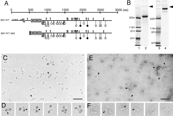
Figure 3.
Expression and electronmicroscopic analysis of the extracellular domain of Ten-m1. (A) Cartoon of Ten-m1 and the secreted COOH-terminal part of Ten-m1 (Ten-m1sec). (B) Coomassie blue staining of molecular mass standards (lanes 1 and 3) and purified Ten-m1sec (lanes 2 and 4) separated by 5% SDS-PAGE under reducing (lanes 1 and 2) and nonreducing (lanes 3 and 4) conditions. The arrowhead indicates the beginning of the separating gel. (C–F) Electron micrographs after glycerol spraying/rotary shadowing (C and D) and negative staining (E and F) of Ten-m1sec. Representative fields of molecules (C and E) and selected species of the same material (D and F) are shown. Pairs of spherical domains, connected by thin elongated rods, are visible. Some of the spheres are resolved into three globular subdomains as indicated by arrowheads (D and F). Arrowheads (C and E) also indicate pairs of Ten-m1sec dimers interacting with each other. Bars, 50 nm.

