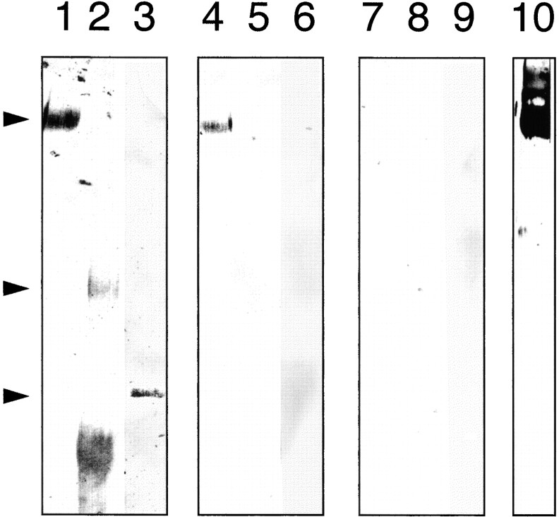
Figure 9.
Homophilic interaction of Ten-m1. Western blot of the recombinant extracellular domain of Ten-m1 (lanes 1, 4, 7, and 10), recombinant neurocan core protein (lanes 2, 5, and 8), and BSA (lanes 3, 6, and 9), separated on a 5% (Ten-m1 and neurocan) or on a 7% (BSA) SDS-PAGE performed under nonreducing conditions. Lanes 1–3 were stained with amido black. Arrowheads indicate the 400-kD Ten-m1sec dimer, the 250-kD neurocan core protein, and the 67-kD BSA band. Lanes 4–6 were incubated with purified AP-ten-m1 fusion protein, and lanes 7–9 with purified AP alone. Lane 10 was incubated with the purified anti–Ten-m1 antiserum and AP-conjugated secondary antibodies.

