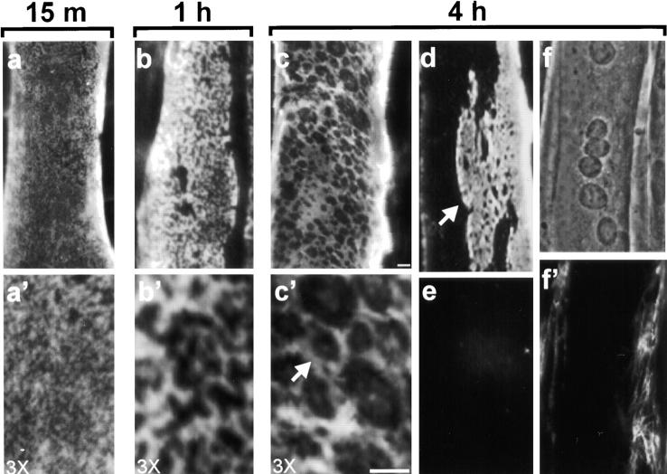
Figure 1.
Laminin binds to the myotube surface and forms a network. Laminin attached to the surface of C2C12 myotubes was visualized using polyclonal antibodies against specific laminin subunits followed by rhodamine-conjugated secondary antibodies. After incubation with 1 μg/ml laminin-1 for 15 min, laminin was seen on the dorsal myotube surface in a diffuse punctate pattern (a and a′). In 1 h, laminin was organized in a more aggregated, reticular pattern (b and b′). Myotubes incubated with 10 μg/ml laminin-1 (c, c′, and d) for 4 h showed an extensive repeating polygonal network of laminin on their surface (see c′ for a higher magnification view of this network, arrow depicts polygonal unit). At 4 h, many myotubes also contain regions cleared of laminin, with surface networks appearing in more compact clusters (d). Control myotubes incubated with BSA show little laminin staining (e). Fibronectin (10 μg/ml for 4 h) forms fibrillar structures primarily in regions containing myoblasts, not myotubes (f, phase micrograph, f′, antifibronectin). Bar, 5 μm.

