
Figure 5.
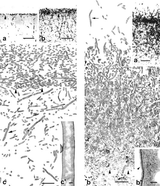
(Left) Vertical sections of colonies of S. antibioticus grown for 12 h (a) and 24 h (b and c). (a and b) Semithin sections showing the general organization of the colonies. Colonies are formed by a thin, compact layer of substrate hyphae that develop above the culture medium and by a large, hyphal network that develops within it. (c) Ultrathin section throughout the substrate mycelium. The hyphae are all similar in ultrastructural appearance (a granular, electron-dense cytoplasm with nucleoids of different size and shape distributed throughout it) and do not display symptoms of cellular disorganization. (c′) Enlarged detail of the hypha pointed to in c (arrow). (a–c) Arrowheads point to the surface of the culture medium. (Right) Vertical sections of a colony of S. antibioticus grown for 36 h. (a) Semithin section showing formation of aerial hyphae in the upper region of the colony. (b) Ultrathin section extending from the upper region of the colony to the surface of the culture medium. Hyphae with evident symptoms of cellular degeneration are visible near the boundary with the culture medium. (b′) Enlarged detail of the hypha pointed to in b (arrow) showing the thin sheath (arrowhead) that surrounds it. Arrowheads in b indicate the agar surface. Bars in left panels: (a and b) 20 μm; (c) 5 μm; (c′) 0.2 μm. Bars in right panels: (a) 15 μm; (b) 5 μm; (b′) 0.1 μm.