
Figure 7.
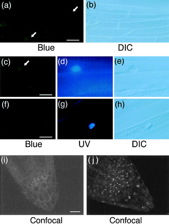
Fluorescence microscopic images of hypocotyl and root tip cells in dark-grown PBG-5 and wild-type seedlings. Hypocotyl specimens were stained with Hoechst No. 33342 and viewed under epifluorescence optics with blue (a, c, and f) and UV (d and g) excitation or under DIC optics (b, e, and h). Root tip specimens were observed on an inverted laser scan microscope (LSM410 invert; Carl Zeiss Jena) with a combination of 488 nm laser excitation and 515 nm longpass emission filter (i and j). Arrows indicate fluorescence detected in the nuclear regions. (a and b) Dark-grown PBG-5 hypocotyl cells, ×40 objective. Bar, 25 μm. (c–e) Dark-grown PBG-5 hypocotyl cells, ×100 objective. Bar, 10 μm. (f–h) Dark-grown wild-type hypocotyl cells, ×100 objective. Bar, 10 μm. (i) Dark-grown PBG-5 root tip cells, ×40 objective. Bar, 20 μm. (j) Light-grown PBG-5 root tip cells, ×40 objective.