
Figure 4.
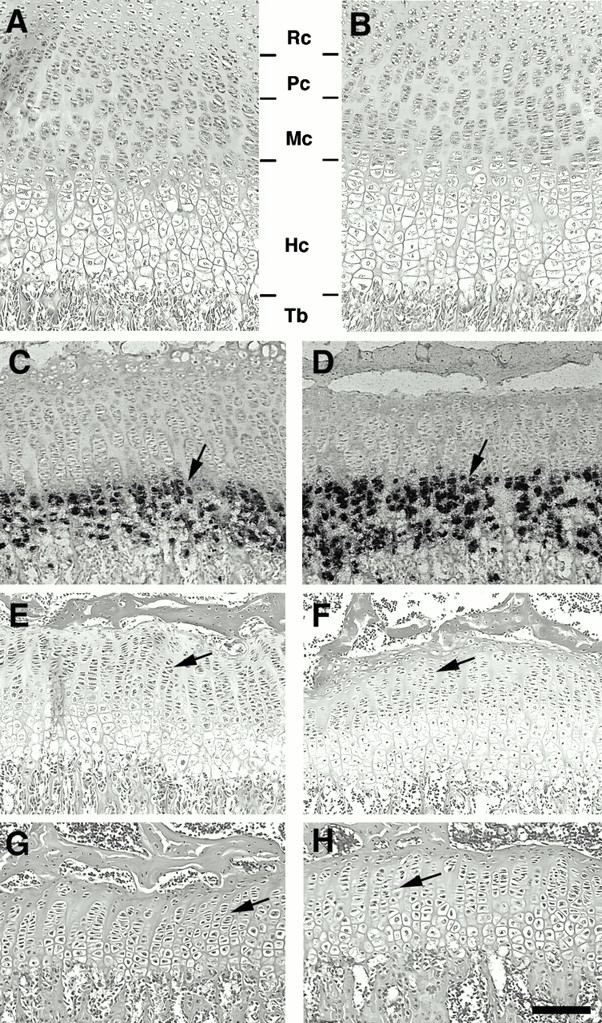
Histologic analysis of epiphyseal growth plates of Smad3 ex8/ex8 mice. (A and B) Sections of wild-type (A) and mutant (B) growth plates from P12 tibiae. Chondrocytes are divided into four distinct zones: resting (Rc), proliferation (Pc), maturing (Mc), and hypertrophic (Hc) chondrocytes. No apparent difference was detected. (C–H) Sections of wild-type (C, E, and G) and mutant (D, F, and H) growth plates isolated from P21, (C and D), P40 (E and F), and P60 (G and H) mice. Arrows in C and D point to hypertrophic chondrocytes positive for type X collagen. Arrows in E and H point to chondrocyte columns. Tb, trabecular bone. Bar: (A, B, G, and H) 80 μm; (C, D, E, and F) 120 μm.