
Figure 3.
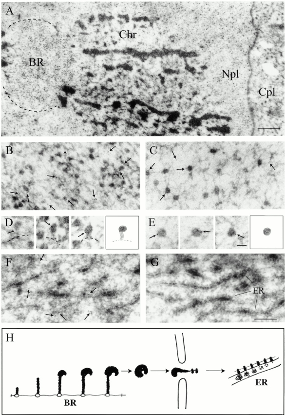
Immunoelectron microscopic localization of actin in C. tentans salivary gland cells. (A) Nucleus in a salivary gland cell. Chr, polytene chromosome; Npl, nucleoplasm; Cpl, cytoplasm. (B) Balbiani ring. (C) Nucleoplasm. (D) Growing BR particles and a schematic drawing. Broken line, putative position of chromatin axis. (E) Nucleoplasmic BR particles and a schematic drawing. (F and G) Cytoplasm. (H) Schematic presentation of the assembly and transport of BR particles. (B–F) Antiactin antibody; (G) control antibody. Gold particles have been marked with small arrows. Bar, 1 μm (A), 200 nm (B, C, F, and G), and 50 nm (D and E).