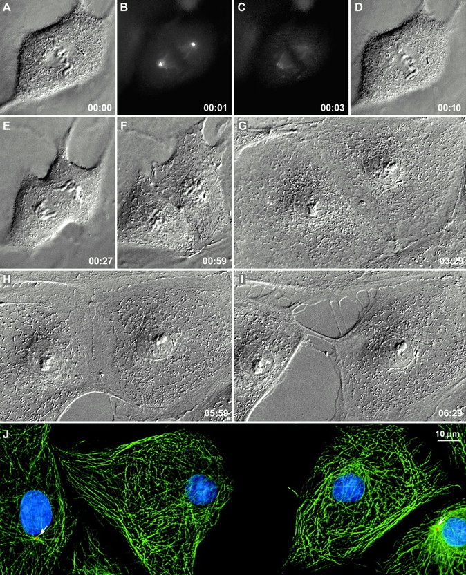
Figure 2.

Cells can divide normally without centrosomes. After destroying both centrosomes during metaphase (B and C), this cell was followed by time-lapse differential interference contrast microscopy. It underwent a normal telophase (E and F) and cytokinesis (E–H), and ∼5 h later the midbody broke as the two daughter cells began to migrate from each other (H and I). At this time, the culture was fixed and stained as described in the legend to Fig. 1. Note that the two acentrosomal daughter cells contained normal numbers of Mts but that, when compared with neighboring centrosomal cells (J, arrows), the Mt lacked a sharp focus. PtKG cells. Time in h:min. The image in I is a maximal intensity projection through the cell volume after deconvolution.
