Figure 5.
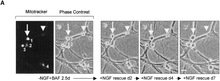
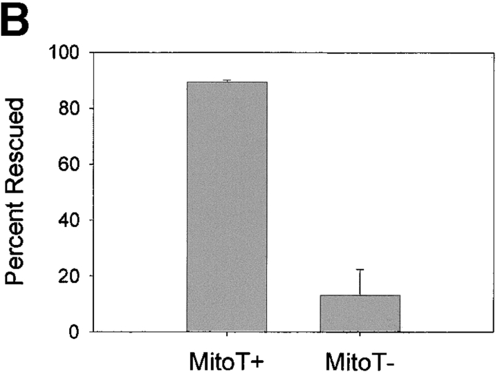
Mitotracker staining predicts rescue with NGF readdition. (A) NGF-maintained sympathetic neurons were deprived of NGF in the presence of 50 μM BAF for 60 h (2.5 d). Cells were loaded with Mitotracker orange (250 nM for 1 h) and photographed to document the status of Mitotracker staining in cells. A representative field shows 4 cells out of 10 that were Mitotracker positive (numbered 1–4; arrow points to one MitoT+ cell). Cells were then treated with NGF, and the same field of cells were photographed after 2, 4, and 7 d of NGF readdition. Photographs show that the Mitotracker positive cells were rescued with NGF readdition, whereas the Mitotracker negative cells were not (arrowhead shows one MitoT– cell). (B) Data showing the correlation between Mitotracker positivity and the ability of cells to be rescued with NGF readdition. Cells were treated as described in A. Results are mean (± range) of two independent experiments with ∼200 cells counted per experiment. Bar, 20 μm.


