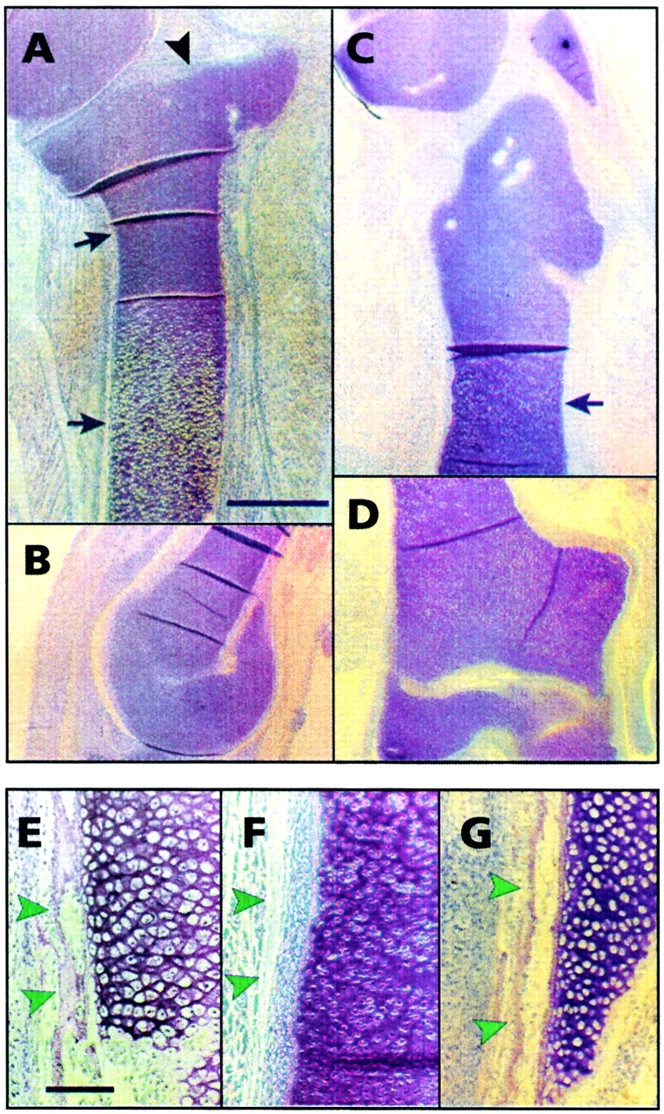
Figure 3.

Functional analyses of C-1-1 and ch-ERG roles in chondrocyte and long bone development. Retroviral particles encoding C-1-1 or ch-ERG were implanted in stage 22–23 chick embryo leg buds in ovo; insert-less retroviral particles were implanted in companion embryos as control. Embryos were reincubated and examined on day 10 by histology. (A and B) Proximal one-third portion and distal epiphysis of control tibiotarsus, respectively. Note the characteristic flat proximal articular epiphysis (A, arrowhead) and the round distal epiphysis (B), and the long metaphyseal shaft with chondrocytes undergoing maturation and hypertrophy (arrows in A). (C and D) Entire C-1-1 virus-infected tibiotarsus. These pictures were taken at the same magnification as (A and B) and thus show that the C-1-1 tibiotarsus is nearly half the length of the control. Both proximal and distal epiphyses are severely malformed, and the metaphyseal-diaphyseal shaft (arrow in C) lacks a normal growth plate and hypertrophic chondrocytes. (E) Diaphyseal portion of control tibiotarsus viewed at higher magnification; it displays mineralizing post-hypertrophic chondrocytes, invading bone and marrow precursor cells, and a well-developed intramembranous bone collar (arrowheads). (F) Diaphyseal portion of C-1-1 tibiotarsus in which hypertrophic chondrocytes, marrow invasion and a bone collar (arrowheads) are all absent. (G) Diaphyseal portion of ch-ERG virus-infected tibiotarsus. Note that it closely resembles control tibiotarsus and contains hypertrophic chondrocytes and a bone collar (arrowheads). Bars: (A–D) 1.5 mm; (E–G) 60 μm.
