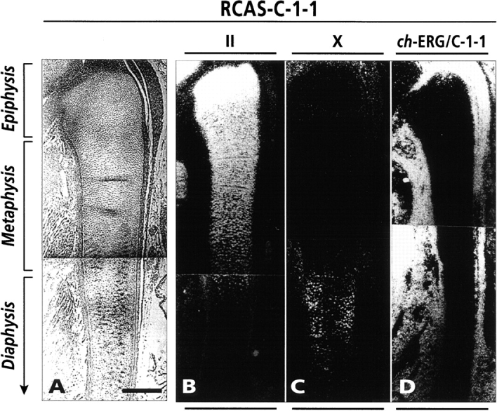
Figure 5.

In situ hybridization analysis of gene expression in day 10 chick embryo fibula. Longitudinal sections of fibula and surrounding tissues from C-1-1 virus-infected embryos were processed for in situ hybridization with indicated probes. (A) Phase micrograph; (B) type II collagen hybridization, showing a characteristic strong signal in epiphysis and metaphysis and a much reduced signal in hypertrophic diaphysis; (C) type X collagen hybridization, showing strong signal in hypertrophic diaphysis; and (D) hybridization with common ch-ERG/C-1-1 probe, showing lack of signal in fibula but extremely strong signal in surrounding perichondrial and connective tissues. Bar, 200 μm.
