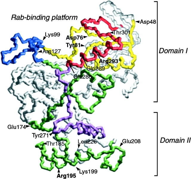
Figure 1.
Potential location of substitutions (yeast numbering) based on the crystal structure of bovine α-GDI. The tentative location of each of the residues (Mrs6p residue numbering) mutated in this study based on their homologous residue in α-GDI (Table ) are indicated (Schalk et al. 1996). The insert region (blue) and the Rab-binding region containing SCRs 1B (yellow) and 3B (red) are highlighted at the top of GDI and form domain I. The conserved face of GDI containing SCRs 2 (green) and 3A (purple) link domain I with a second domain II, and are oriented to the front of the image (Schalk et al. 1996). Bolded residues are those that have prominent phenotype in altering REP function.

