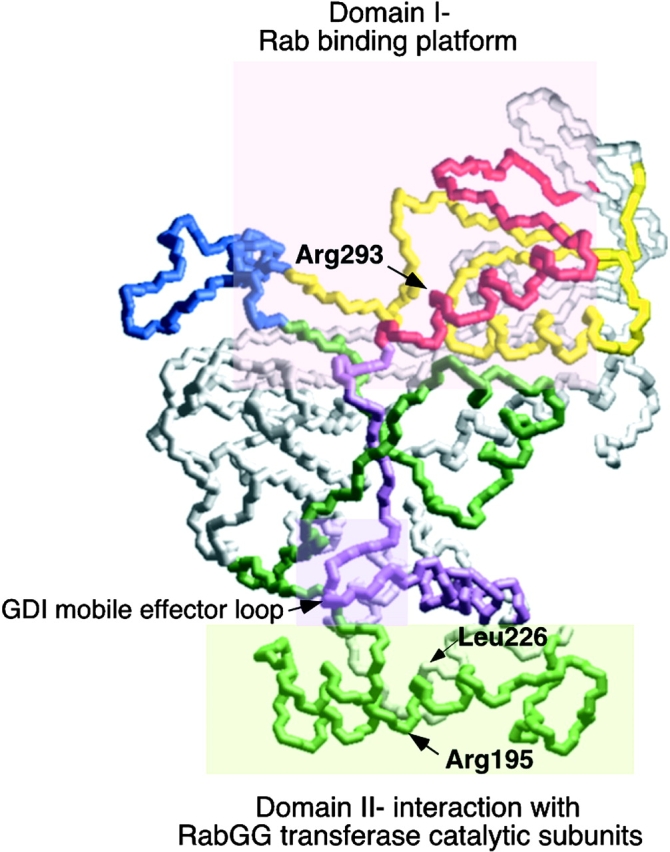
Figure 10.

Domain organization of REP for Rab-binding and prenylation. The figure highlights principle regions involved in REP function based on the homologous residues in GDI through sequence alignment. Residues shaded by the pink box in upper region (Domain I) directs Rab interaction; residues shaded by the light green box in the lower region (Domain II) participate in recognition of the catalytic subunits of GG Tr II. The mobile effector loop involved in recognition of recycling factors by GDI (Luan et al. 2000), but not REP (this study), are indicated by the shaded purple box at the interface of domains I and II.
