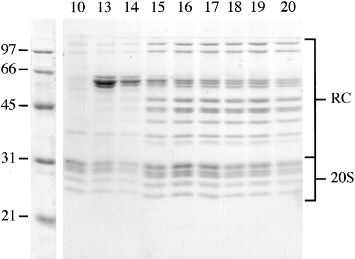
Figure 2.
SDS-PAGE of 26S proteasomes. Fractions 10 and 13–20 from the sucrose density gradient centrifugation were separated on a 12.5% SDS gel and the proteins were stained with Coomassie blue. Molecular weights of the marker proteins are shown on the left. For assignment of bands see Fig. 4. Fraction 10 (∼22% sucrose) contained pure 20S proteasomes, and fractions 13–20 (∼24–28%) displayed the typical pattern of subunit components exhibited by 26S proteasomes. Fractions 13–15 were contaminated with another protein complex (possibly a homologue of GroEL).

