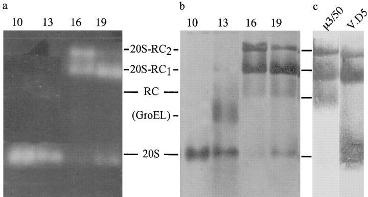
Figure 3.
Nondenaturing PAGE of 26S proteasomes. Fractions 10, 13, 16, and 19 (40 μl) from the sucrose gradients were electrophoresed for 800 V-h on 4.5% native polyacrylamide gels. a, Proteolytic activity of the resolved complexes was detected by fluorogenic peptide overlay with Suc-LLVY-AMC and the proteins were visualized by Coomassie blue stain (b; same gel). c, To further characterize the bands, duplicate samples of fraction 19 were transferred to nitrocellulose membranes and immunostained with μ3/50, an mAb directed against the RC subunit, p39A, and with another, V.D5, directed against the 20S proteasome. The analysis of a, b, and c allowed unambiguous identification of the bands as 20S proteasomes, RCs, 26S proteasomes with only one RC attached (20S-RC1), and 26S proteasomes with two RCs (20S-RC2), as indicated.

