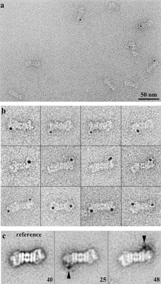
Figure 8.

Mapping of p37A within the RC by EM. a, Electron micrograph of 26S proteasomes incubated with gold-labeled Ub-Al. Bar, 50 nm. b, Gallery of gold-labeled Ub-Al bound to the 26S proteasome after rotational and translational alignment. c, Main classes of unlabeled (reference) and gold-labeled 26S proteasomes after MSA/classification.
