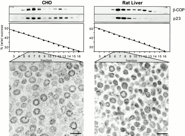
Figure 2.
Characterization of in vitro–generated COPI-coated vesicles. Transport vesicles were generated from either CHO (left) or rat liver (right) Golgi membranes in the presence of bovine brain cytosol, ATP-regenerating system, and GTPγS. COPI vesicles were purified on a continuous sucrose gradient. The gradients were fractionated from the bottom into 18 fractions. Aliquots of fractions 3–16 of the isopycnic gradients were chloroform/methanol-precipitated, and analyzed by running on a 13% SDS-PAGE and Western blotting, using antibodies against β-COP and p23 (top). COPI vesicles are recovered in fractions 6–8, corresponding to an average sucrose concentration of 41% (wt/wt). Below are electron micrographs of COPI-coated vesicles present in fractions 6–8. Bars, 90 nm.

