
Figure 6.
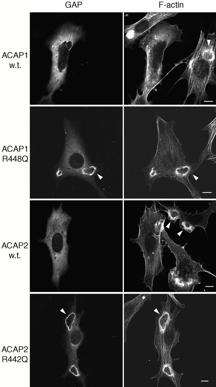
Ectopically expressed ACAPs in PDGF-stimulated NIH 3T3 fibroblasts. NIH 3T3 cells expressing epitope-tagged ACAP1, [R448Q]ACAP1, ACAP2, and [R442Q]ACAP2 were treated for 4 min with PDGF, fixed, and immunostained. Actin was visualized with rhodamine–conjugated phalloidin. Ruffles are indicated by arrowheads. Bars, 10 μm.