
Figure 4.
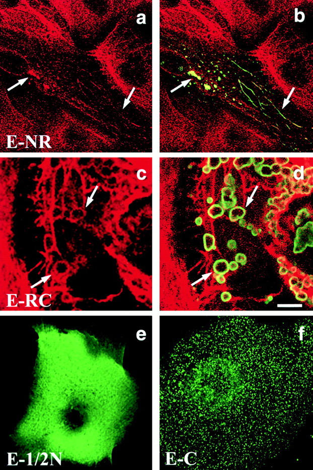
Transient transfection of primary human keratinocytes with different domains of envoplakin. E-NR (a and b); E-RC (c and d); E-1/2N (e); E-C (f). Green fluorescence: anti-FLAG. Red fluorescence: antikeratin (LP34). The same fields of cells are shown in a and b and in c and d. Arrows in a–d show the positions of individual envoplakin aggregates and associated intermediate filaments. Bar: (a and b) 9.3 μm; (c and d) 5 μm; (e) 4 μm; (f) 4.4 μm.