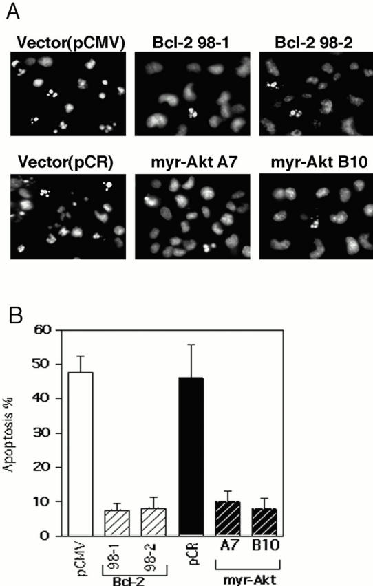
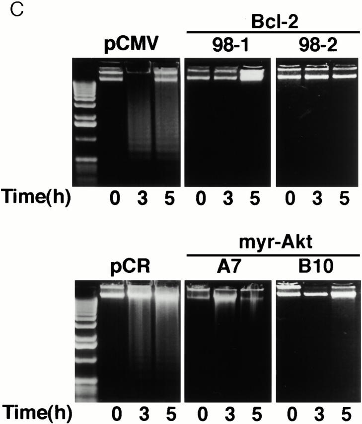
Figure 4.
Inhibition of apoptosis by constitutively active Akt and Bcl-2. (A) Fluorescence photomicrographs of Hoechst 33342 staining of HMN1 cells transfected with pCMV, human Bcl-2, pCR3.1, or myr-AktΔ4-129. Cells were treated with 30 μM C2-ceramide for 5 h. (B) Constitutively active Akt and Bcl-2 inhibit ceramide-induced HMN1 cell apoptosis. Cells were treated with 30 μM C2-ceramide for 5 h and apoptosis was assayed by Hoechst 33342 staining. Data represent mean ± SEM for three independent experiments. (C) Constitutively active Akt and Bcl-2 inhibit DNA fragmentation in HMN1 cells. Cells were treated with 30 μM C2-ceramide for 0, 3, or 5 h, and soluble DNA was extracted for DNA fragmentation assay. Data are from a representative experiment performed three times.