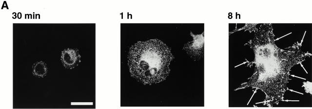
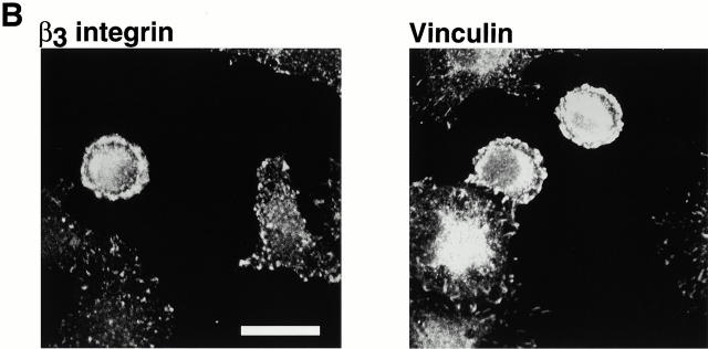
Figure 1.
Distribution of β3 integrin and vinculin in spreading BAE cells. BAE cells were allowed to spread on fibronectin-coated coverslips for 30 min, 1 h, or 8 h (A) or on vitronectin-coated coverslips for 30 min (B). Cells were fixed and permeabilized. Cells were stained with antibodies against β3 integrin or vinculin. Arrows in A indicate focal adhesions. Bars, 20 μm.


