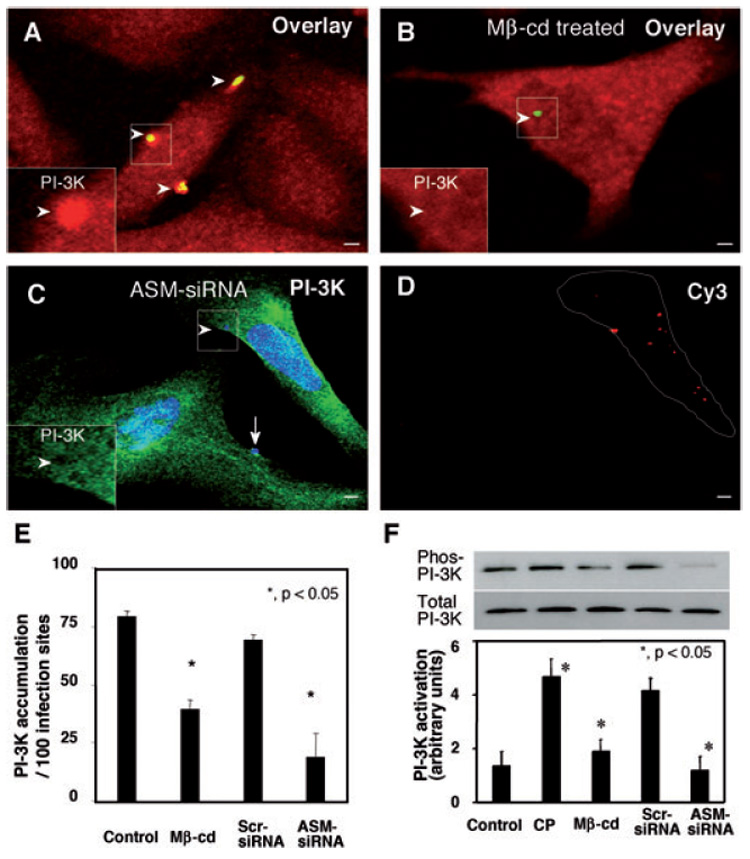
Fig. 6. Inhibition of SEM aggregation hinders C. parvum-induced accumulation and activation of PI-3K in infected cells.

A–D. H69 cells were treated with 10 mM methyl-β-cyclodextrin (Mβ-cd) or ASM-siRNA and then infected with C. parvum sporozoites. Accumulation and activation of PI-3K were determined by dual immunofluorescent labelling and Western blot analysis of tyrosine-phosphorylated p85 subunit of class IA PI-3K as we previously reported (Chen et al., 2004a). The left lower insets in (A)–(C) show labelling of PI-3K in the boxed regions. Whereas non-treated cells showed strong accumulation of PI-3K at infection sites [arrowheads in (A) and inset], treatment of cells with Mβ-cd reduced accumulation of PI-3K [arrowhead in (B) and inset]. Inhibition of PI-3K accumulation was also detected in cells transfected with Cy3-tagged ASM-siRNA [arrowheads in (C) and inset]. In contrast, non-transfected cells showed strong PI-3K accumulation [arrow in (C)]. Cells transfected with ASM-siRNA were identified by positive fluorescence of Cy3 [as outlined in (D)]. Bar = 5 µm.
E. Quantitative analysis of PI-3K accumulation by dual immunofluorescent labelling.
F. Activation of PI-3K as assessed by Western blot analysis of tyrosine-phosphorylated p85 subunit of PI-3K. The PI-3K p85 subunit was immunoprecipitated from whole-cell lysates, separated by SDS-PAGE and then probed for phosphotyrosine. The same whole-cell lysates were also probed for p85 to demonstrate unchanged total p85 protein level. *P < 0.05, compared with control. CP, C. parvum; Scr-siRNA, scrambled siRNA.
