
Fig 4. δPKC modulates the ER stress response of the myocardium in a model of cardiac ischemia and reperfusion injury.
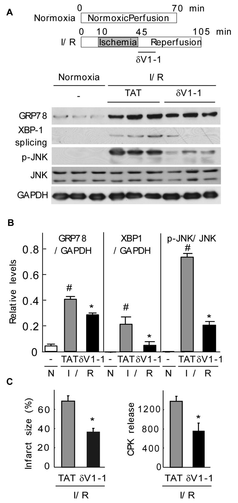
A. Normoxic control hearts and hearts that underwent ischemia and reperfusion were homogenized and total extracts were isolated. The levels of GRP78, spliced XBP1 and phospho-JNK were determined by Western blot.
B. Quantitative data of the hearts described in (A). Values represent mean ± S.E. of three animals in each group (N: normoxia; I/R: ischemia/reperfusion). Student t test; * p<0.05 vs. TAT treatment, # p< 0.05 vs. control.
C. Hearts were subjected to ischemia-reperfusion and treated at the onset of reperfusion with TAT control peptide or δV1-1 and the infarct size (left panel) and cell survival, as demonstrated by the decrease in CPK release (right panel), were determined. Data are expressed as mean ± S.E. of three animals in each group. Student t test; * p<0.05 vs. TAT treatment.