
Figure 5.
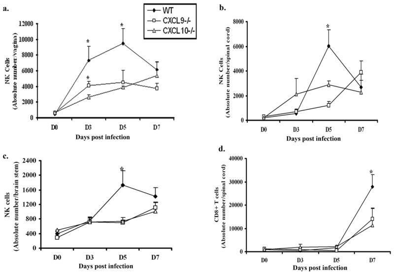
NK and CD8+ T cells infiltration into infected tissue of chemokine knockout mice is reduced or delayed. WT, CXCL9−/− and CXCL10−/− mice (n= 6/group) were infected with HSV-2 (2000 pfu/vagina) and subsequently exsanguinated at indicated times post infection (pi) and (a) vaginal tissue, (b) spinal cord, and (c) brain stem samples were processed and analyzed for NK cell (NK1.1+CD3−CD45high) content by flow cytometry. (d) Similarly, spinal cords were processed and analyzed for CD8+ T (CD3+CD8+CD45high) cells content using flow cytometry. Day 0 time point represents uninfected controls. Each point represents the mean ± SEM summarizing the results of three independent experiments. *, p<0.05 comparing WT to CXCL9−/− and CXCL10−/−.