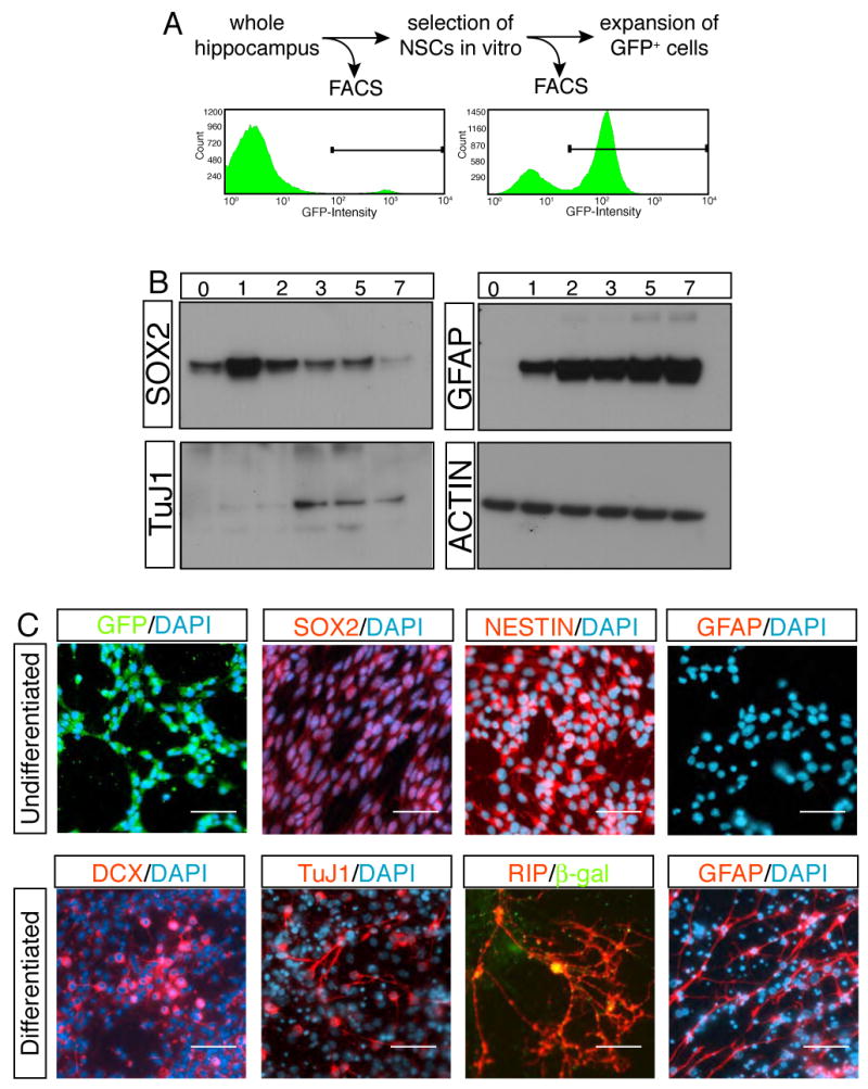
Figure 2. Sox2-GFP cells are the origin of in vitro NSCs.

The proliferation capacity of Sox2-GFP cells was measured by FACS analysis. Sox2-GFP cells, which comprised only 6% of the total hippocampus cells, survived and expanded, forming the majority of colonies in vitro (A). Western analysis showed the progress of differentiation in time course (B). The expressions of neuronal (TuJ1) and glial markers (GFAP) were associated with the down-regulation of SOX2. Immunocytochemistry revealed the differentiation of in vitro cultured Sox2-GFP cells to neural lineages (C). Note that co-culture paradigm with primary neurons was used for differentiation of Sox2-GFP cells to oligodendrocytes (RIP+ cells). Legends in (B); 0, one day after plating cells but still culturing in FGF2, EGF containing growth medium; 1-7, 1 to 7 days after substituting growth medium with Forskolin-containing differentiation medium. Scale bar: 50 μm.
