
Fig. 2. MCP-1-EGFP localizes to large dense-core vesicles (LDCVs) in DRG neurons.
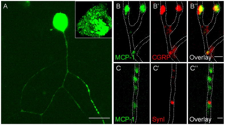
Cultured DRG neurons were infected with an MCP-1-EGFP expressing adenovirus and stained for CGRP and SynI. A) MCP-1-EGFP was concentrated in the perinuclear area, and localized in a punctate pattern in the soma and along axons. Signals in the soma were saturated to allow appreciation of the punctate localization along axons. An unsaturated image of a soma is magnified in the inset. B-C) Magnified view of axonal localization of MCP-1-EGFP. B-B’’) MCP-1-EGFP co-localized with CGRP. C-C’’) MCP-1-EGFP did not co-localize with SynI. Scale bars: A, 20 μm; B’’, C’’, 1 μm.