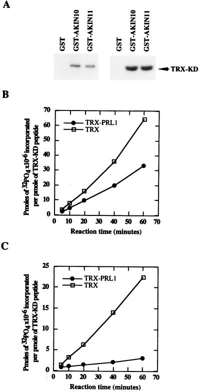
Figure 3.
Autophosphorylation, substrate specificity, and inhibition of AKIN10 and AKIN11 by PRL1 in vitro. (A Left) GST-AKIN10 and GST-AKIN11 on glutathione-Sepharose were incubated with [γ-32P]ATP. After elution and SDS/PAGE separation, the autophosphorylation was detected by autoradiography. (Right) Phosphorylation of the TRX-KD substrate by immobilized GST-AKIN10 and GST-AKIN11 was detected by SDS/PAGE followed by autoradiography. (B) Immobilized GST-AKIN10 (1 μg) was preincubated for 30 min with either 1 μg TRX-PRL1 (●) or 1 μg His6-thioredoxin (□) protein before the addition of the TRX-KD substrate and [γ-32P]ATP. Equal amounts of samples withdrawn at different time points were separated by SDS/PAGE, and the phosphorylated TRX-KD bands were excised to measure the incorporated radioactivity. (C) PRL1-inhibition assay with GST-AKIN11 (as described in B). Phosphate incorporation per picomole of TRX-KD substrate is plotted against the reaction time.

