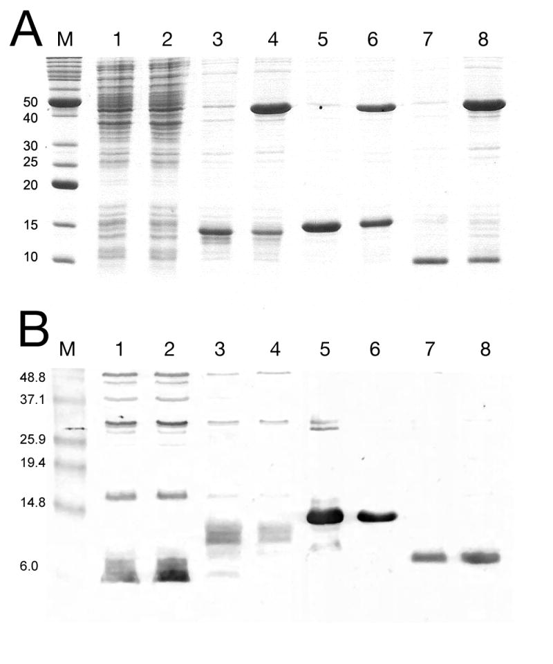
Figure 6.

SDS-PAGE analysis of protein expression from truncated gpO clones, by Coomassie staining (A) or immunological detection with an anti-gpO antibody (B). Lane 1, O* alone; lane 2, O* and gpN; lane 3, O(26–141); lane 4, O(26–141)+gpN; lane 5, O(142–284); lane 6, O(142–284)+gpN; lane 7, O(195–284); and lane 8, O(195–284)+gpN. The truncated gpO proteins are indicated by arrows. M, markers, MW (kDa) as indicated. A few non-specific bands are seen on the immunoblot for the O* and O(26–142) expressions. Also note that the antibody is reportedly less sensitive to the N-terminal half of the protein than to the C-terminal half (Marvik et al., 1994a).
