
Figure 1.
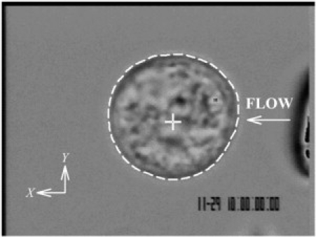
Digital micrograph of a freshly isolated human leukocyte as used in the current experiments. The cell outline is indicated by a white dashed line, and the computed cell centroid is shown as a white cross. The tip of an adjacent pipette used to apply FSS is just visible to the right of the image with the direction of fluid discharge indicated by the arrow.