
Figure 5.
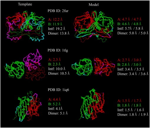
Representative examples of M-TASSER models compared to the best initial template. The left-hand column is the best template superimposed onto the native structure whereas the right-hand column shows the final model superimposed onto the native structure. The thin lines are native structures with monomer chain A colored in red and chain B colored in green. The thick lines are initial templates or final models with residues within 5 Å from native colored in red (chain A) or green (chain B). Residues that lie beyond this distance are in magenta (chain A) or cyan (chain B). For models, the numbers on the left are the RMSD to native over the same aligned residues as the best template; the numbers on the right are the RMSD to native of the entire chain.