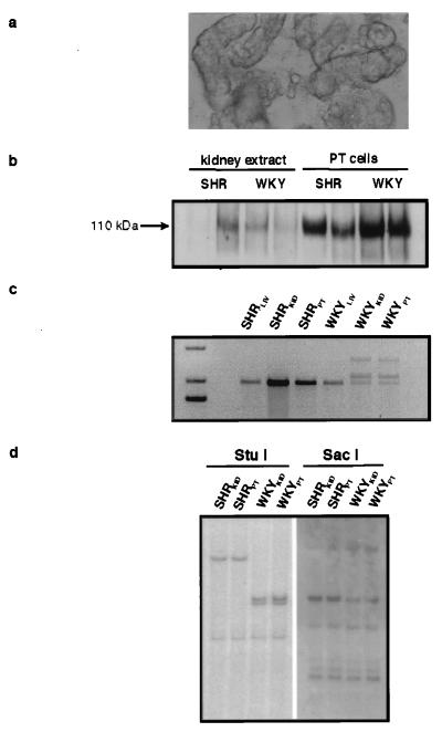
Figure 6.
Analysis of isolated proximal tubular segments. (a) Phase contrast microscopy (×400) of isolated segments showing a homogeneous population of large diameter segments with morphology typical of PT cells with granular cytoplasm. (b) Western blot of protein extracted from either whole kidney (lanes 1–4) or purified PT segments (lanes 5–8) from SHR (lanes 1, 2, 5, and 6) and WKY (lanes 3, 4, 7, and 8) rats probed for presence of the specific PT protein NBC3. (c) RT-PCR analysis of SA mRNA from liver (LIV), kidney (KID), and isolated PT segments (PT) of SHR and WKY. Note the presence of additional PCR products showing exon repetition in RNAs from WKY kidney (WKYKID) and WKY PT cells (WKYPT) only. (d) StuI and SacI restriction analysis of SA gene structure in whole kidney and PT segments of SHR and WKY excluding somatic rearrangement of the SA gene in WKY PT cells.

