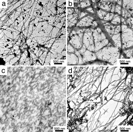
Fig. 3.
Electron micrographs of oligomeric forms of huPrP N–terminal fragments. (a) Fibrils of huPrP23–144 after 4 h of incubation. (b) Bundled fibrils of huPrP23–144 after 12 h of incubation. (c) Aggregates formed by huPrP23–139 after 60 h of incubation. (d) Fibrils of huPrP23–141 after4hof incubation. All proteins were incubated at a concentration of 400 μM in 50 mM potassium phosphate buffer, pH 6.5, at 25°C.

